A small mass can be associated with big problems.
- Molly Benoît

- Jun 10
- 4 min read
Do you feel a small lump on your pet's body, and are you worried about it?
That's perfectly normal.
Unfortunately, it's impossible to tell with the naked eye whether this mass is benign, inflammatory, or cancerous. A mass can sometimes simply be a subcutaneous fatty deposit, but it can also represent a tumorous process that requires prompt medical attention.
Don't wait to investigate the mass you've just identified if you care about your baby's health. In many surgical cases, we observe a significant delay between when the mass is noticed and when steps are taken to determine its nature. However, the larger a mass grows, the more complex its removal will be and the more limited the surgical options will be, which will have a considerable impact on the patient's prognosis.
The appearance of a mass is not sufficient for a reliable diagnosis. Two masses with a very similar appearance can be composed of completely different cells and have distinct biological behaviors. This is why it is essential not to rely solely on its appearance to assess its severity.
Let's play a game to better illustrate this concept. Of these four photos, which of these masses do you think is a mast cell tumor, a type of cancerous skin mass?
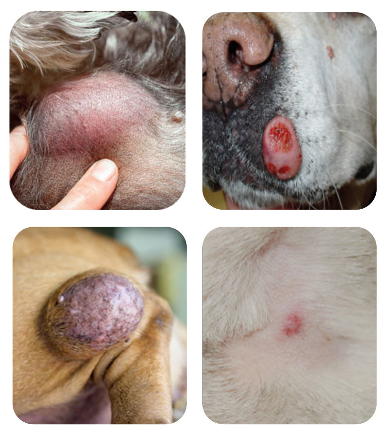
If you answered that they all are, you are absolutely right! Appearance, size and/or location do not allow us to determine the type of mass.
So, how can we determine whether the mass effect you have identified on your animal is dangerous to its health or not?
First diagnostic step: cytology or fine needle aspiration
Cytology of a mass is a simple test that involves taking a few cells with a small needle. The sample is usually taken anesthetically, but may require light sedation depending on the location of the mass and the patient's responsiveness.
The cells are then spread on a slide and examined under a microscope by a veterinarian or pathologist. This test often provides a quick initial indication of the mass's nature: inflammation, infection, cyst, benign or malignant mass. In many cases, cytology also helps guide the next diagnostic and treatment steps.
Cytology provides a diagnostic result in 80% of cases. Factors influencing the success of a diagnostic result include the type of mass, its location, and the quality of the sample. For cutaneous or subcutaneous masses in dogs and cats, one study showed that cytology is a reliable tool compared to biopsy and histopathological analysis, which remains the gold standard. Some studies also report very high accuracy rates for certain masses or locations, sometimes exceeding 90%, but this is not guaranteed for all masses.
It is important to know that cytology does not always provide a definitive answer. Sometimes the sample does not contain enough cells, the mass bleeds excessively, contaminating the sample, or the tumor type is not clearly identifiable by cytology. In these situations, a biopsy or removal of the mass with laboratory analysis may be recommended to obtain a more precise diagnosis.
Second diagnostic step: the biopsy
When cytology is inconclusive, a biopsy may be recommended to obtain more information about the nature of the mass. This procedure involves surgically removing a small fragment of the mass, which is then sent to a laboratory for analysis. Because the sample is larger and, more importantly, better organized than with a fine-needle aspiration, it often allows for a more precise diagnosis.
In some cases, however, a biopsy is not necessary or is not the preferred option. The decision depends on several factors, including the location of the mass, its size, its appearance, and the level of clinical suspicion. If the diagnosis already seems clear enough or a surgical plan can be established immediately, your veterinarian may recommend proceeding directly to staging and/or surgery.
To learn more about staging, consult our information sheet on cutaneous and subcutaneous masses:
The treatment: surgery
This is where our team comes in. Once the origin of the mass is determined, we can advise you on the best surgical approach for its removal. It's important to understand that a benign mass is not removed in the same way as a malignant one. Therefore, the surgical plan must always be tailored to the type of mass suspected or diagnosed.
When a mass is considered cancerous, it is essential to plan adequate surgical margins, meaning that not only the visible mass is removed, but also a portion of surrounding healthy tissue. The goal is to minimize the risk of leaving microscopic tumor cells in place, which could promote recurrence after surgery.
The following photos clearly illustrate the importance of acting quickly when a mass appears. The larger a mass becomes, the more complex the surgery will be. Wider margins are often necessary, which increases the size of the incision, the tension on the surgical site, and, in some cases, the complexity of the reconstruction. Conversely, a small mass treated early often allows for a simpler procedure under better conditions.

At Coupez, we are always available to answer your questions. You can also schedule a virtual consultation with our surgeons to discuss the best way to manage your pet's weight by clicking here:
Our surgeons will assist you in assessing the mass and in developing the diagnostic and surgical plan best suited to your animal.


